International T1 Multicentre
|

|
Overview |
Aims |
|
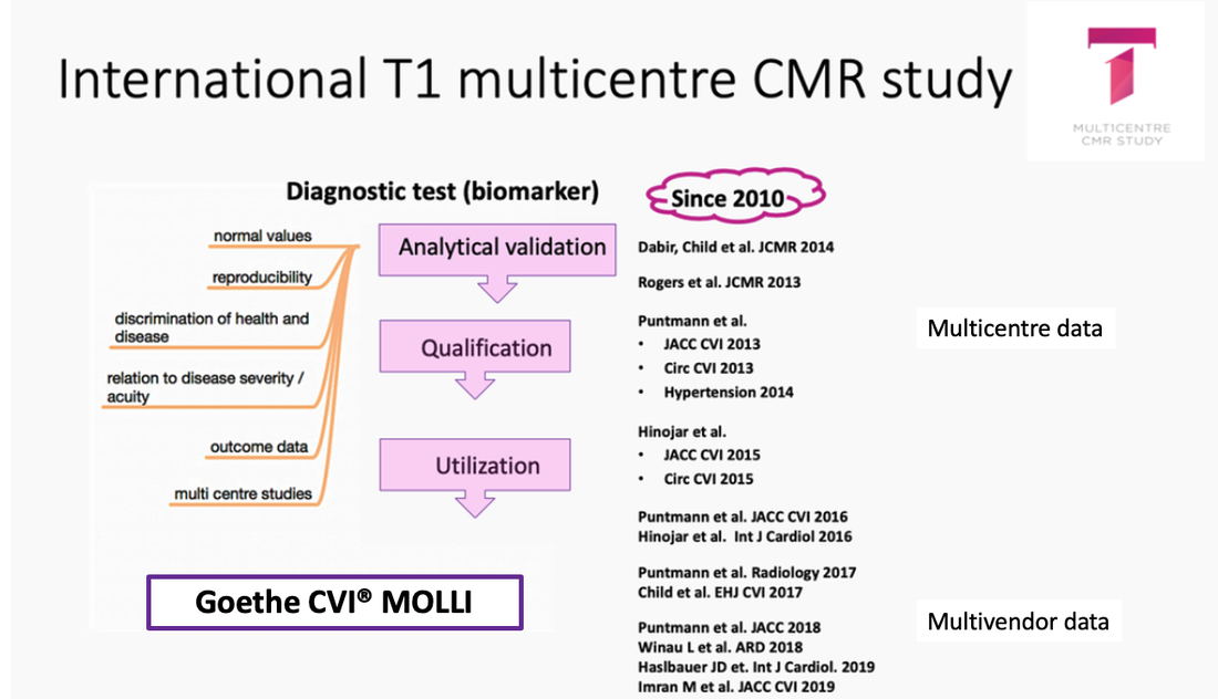
The international T1 mapping multicentre consortium was established in 2012 with aim to accumulate a large data collective in a number of disease contexts using a unified T1 mapping sequence based on modified Look-Locker Imaging (MOLLI) and postprocessing approach (ConSept). The consortium originally consisted of CMR centres using 1.5 and 3T Philips scanners that adopted standardised T1-mapping MOLLI sequence, which was developed and validated by Drs Nagel and Puntmann. Since 2016, this sequence is also available on Siemens 1.5T and 3.0T scanners, thus, providing the first T1 mapping sequence, which is transferable cross-vendor in terms of normal values and clinical experience. The respective sequences, sequence parameters, normal values and clinical evidence have been published in a number of publications (below) and are freely downloadable from this website.
|
Biomarker qualification pathway of T1 mapping in characterisation of diffuse myocardial disease:
Registrationwww.clinicaltrials.gov (NCT02407197, NCT03749343)
|
|
Why T1 mapping
|
Link to the video: Puntmann VO: T1 mapping is ready for prime time - PRO (EuroCMR 2016)
Inclusion Criteria |
Exclusion Criteria |
|
1. Verified MR unsafe devices or objects
|
Endpoints |
Primary outcome
1. All cause mortality at 2 and 5 years |
Secondary outcome
1. Cardiovascular mortality at 2 and 5 years 2. Heart failure endpoint (HF death and HF Hospitalization) at 2 and 5y |
Participating sites |
T1 and T2 mapping sequences |
|
1. Goethe CVI, Frankfurt, Germany
2. Kerckhoff Klinik, Bad Nauheim, Germany. 3. University Hospital La Paz, Madrid, Spain 4. Guys and St Thomas' NHS Trust, London UK 5. St Vincent University Hospital, Sydney, Australia 6. German Heart Institute Berlin, Berlin, Germany 7. University Hospital Mainz, Germany |
IMPORTANT INFORMATION regarding MOLLI sequences: there are several variants of MOLLI sequences available. These sequences are not identical, as they rely on very different imaging parameters. Consequently, they have very different normal values and have different sensitivity/specificity for disease detection. The MOLLI sequences are not interchangeable.
|
Publications
Validation:
1. Child N, Suna G, Dabir D, Yap ML, Rogers T, Kathirgamanathan M, Arroyo-Ucar E, Hinojar R, Mahmoud I, Young C, Wendler O, Mayr M, Sandhu B, Morton G, Muhly-Reinholz M, Dimmeler S, Nagel E, Puntmann VO. Comparison of MOLLI, shMOLLLI, and SASHA in discrimination between health and disease and relationship with histologically derived collagen volume fraction. Eur Heart J Cardiovasc Imaging. 2017 Dec 11.
2. Puntmann VO, Arroyo Ucar E, Hinojar Baydes R, Ngah NB, Kuo YS, Dabir D, Macmillan A, Cummins C, Higgins DM, Gaddum N, Chowienczyk P, Plein S, Carr-White G, Nagel E. Aortic stiffness and interstitial myocardial fibrosis by native T1 are independently associated with left ventricular remodeling in patients with dilated cardiomyopathy. Hypertension. 2014 Oct;64(4):762-8.
Standardisation:
3. Rogers T, Dabir D, Mahmoud I, Voigt T, Schaeffter T, Nagel E, Puntmann VO. Standardization of T1 measurements with MOLLI in differentiation between health and disease--the ConSept study. J Cardiovasc Magn Reson. 2013 Sep 11;15:78.
Normal Values:
4. Dabir D, Child N, Kalra A, Rogers T, Gebker R, Jabbour A, Plein S, Yu CY, Otton J, Kidambi A, McDiarmid A, Broadbent D, Higgins DM, Schnackenburg B, Foote L, Cummins C, Nagel E, Puntmann VO. Reference values for healthy human myocardium using a T1 mapping methodology: results from the International T1 Multicenter cardiovascular magnetic resonance study. J Cardiovasc Magn Reson. 2014 Oct 21;16:69.
Proof of concept studies:
5. Puntmann VO, Voigt T, Chen Z, Mayr M, Karim R, Rhode K, Pastor A, Carr-White G, Razavi R, Schaeffter T, Nagel E. Native T1 mapping in differentiation of normal myocardium from diffuse disease in hypertrophic and dilated cardiomyopathy. JACC Cardiovasc Imaging. 2013 Apr;6(4):475-84.
6. Puntmann VO, D'Cruz D, Smith Z, Pastor A, Choong P, Voigt T, Carr-White G, Sangle S, Schaeffter T, Nagel E. Native myocardial T1 mapping by cardiovascular magnetic resonance imaging in subclinical cardiomyopathy in patients with systemic lupus erythematosus. Circ Cardiovasc Imaging. 2013 Mar 1;6(2):295-301.
7. Hinojar R, Foote L, Arroyo Ucar E, Jackson T, Jabbour A, Yu CY, McCrohon J, Higgins DM, Carr-White G, Mayr M, Nagel E, Puntmann VO. Native T1 in discrimination of acute and convalescent stages in patients with clinical diagnosis of myocarditis: a proposed diagnostic algorithm using CMR. JACC Cardiovasc Imaging. 2015 Jan;8(1):37-46.
8. Hinojar R, Varma N, Child N, Goodman B, Jabbour A, Yu CY, Gebker R, Doltra A, Kelle S, Khan S, Rogers T, Arroyo Ucar E, Cummins C, Carr-White G, Nagel E, Puntmann VO. T1 Mapping in Discrimination of Hypertrophic Phenotypes: Hypertensive Heart Disease and Hypertrophic Cardiomyopathy: Findings From the International T1 Multicenter Cardiovascular Magnetic Resonance Study. Circ Cardiovasc Imaging. 2015 Dec;8(12).
9. Isted A, Grigoratos C, Bratis K, Carr-White G, Nagel E, Puntmann VO. Native T1 in deciphering the reversible myocardial inflammation in cardiac sarcoidosis with anti-inflammatory treatment. Int J Cardiol. 2016 Jan 15;203:459-62.
10.Hinojar R, Varma N, Child N, Goodman B, Jabbour A, Yu CY, Gebker R, Doltra A, Kelle S, Khan S, Rogers T, Arroyo Ucar E, Cummins C, Carr-White G, Nagel E, Puntmann VO. T1 Mapping in Discrimination of Hypertrophic Phenotypes: Hypertensive Heart Disease and Hypertrophic Cardiomyopathy: Findings From the International T1 Multicenter Cardiovascular Magnetic Resonance Study. Circ Cardiovasc Imaging. 2015 Dec;8(12).
11. Hinojar R, Foote L, Sangle S, Marber M, Mayr M, Carr-White G, D'Cruz D, Nagel E, Puntmann VO. Native T1 and T2 mapping by CMR in lupus myocarditis: Disease recognition and response to treatment. Int J Cardiol. 2016 Nov 1;222:717-26
12. Puntmann VO, Isted A, Hinojar R, Foote L, Carr-White G, Nagel E. T1 and T2 Mapping in Recognition of Early Cardiac Involvement in Systemic Sarcoidosis. Radiology. 2017 Apr 27:162732.
13. Winau L, Hinojar Baydes R, Braner A, Drott U, Burkhardt H, Sangle S, D'Cruz DP, Carr-White G, Marber M, Schnoes K, Arendt C, Klingel K, Vogl TJ, Zeiher AM, Nagel E, Puntmann VO. High-sensitive troponin is associated with subclinical imaging biosignature of inflammatory cardiovascular involvement in systemic lupus erythematosus. Ann Rheum Dis. 2018 Nov;77(11):1590-1598.
14. Haslbauer JD, Lindner S, Valbuena-Lopez S, Zainal H, Zhou H, D'Angelo T, Pathan F, Arendt CA, Bug G, Serve H, Vogl TJ, Zeiher AM, Carr-White G, Nagel E, Puntmann VO. CMR imaging biosignature of cardiac involvement due to cancer-related treatment by T1 and T2 mapping. Int J Cardiol. 2019 Jan 15;275:179-186.
15. Imran M, Wang L, McCrohon J, Yu C, Holloway C, Otton J, Huang J, Stehning C, Moffat KJ, Ross J, Puntmann VO, Vassiliou VS, Prasad S, Kotlyar E, Keogh A, Hayward C, Macdonald P, Jabbour A. Native T1 Mapping in the Diagnosis of Cardiac Allograft Rejection: A Prospective Histologically Validated Study. JACC Cardiovasc Imaging. 2019 Jan 9.
16. Saunders LC, Johns CS, Stewart NJ, Oram CJE, Capener DA, Puntmann VO, Elliot CA, Condliffe RC, Kiely DG, Graves MJ, Wild JM, Swift AJ.
Diagnostic and prognostic significance of cardiovascular magnetic resonance native myocardial T1 mapping in patients with pulmonary hypertension. J Cardiovasc Magn Reson. 2018 Dec 3;20(1):78. doi: 10.1186/s12968-018-0501-8.
17. Chen M, Arcari L, Engel J, Freiwald T, Platschek S, Zhou H, Zainal H, Buettner S, Zeiher AM, Geiger H, Hauser I, Nagel E, Puntmann VO. Aortic stiffness is independently associated with interstitial myocardial fibrosis by native T1 and accelerated in the presence of chronic kidney disease. Int J Cardiol Heart Vasc. 2019 Jun 26;24:100389. doi: 10.1016/j.ijcha.2019.100389.
18. Heinke R, Pathan F, Le M, D'Angelo T, Winau L, Arendt C, Vogl TJ, Zeiher A, Nagel E, Puntmann VO. Towards standardized postprocessing of global longitudinal strain by feature tracking - OptiStrain CMR-FT study. BMC Cardiovasc Disord. 2019 Nov 27;19(1):267. doi: 10.1186/s12872-019-1255-4.
19. Pathan F, Zainal Abidin HA, Vo QH, Zhou H, D'Angelo T, Elen E, Negishi K, Puntmann VO, Marwick TH, Nagel E. Left atrial strain: a multi-modality, multi-vendor comparison study. Eur Heart J Cardiovasc Imaging. 2021 Jan 1;22(1):102-110. doi: 10.1093/ehjci/jez303.
20. Le MTP, Zarinabad N, D'Angelo T, Mia I, Heinke R, Vogl TJ, Zeiher A, Nagel E, Puntmann VO. Sub-segmental quantification of single (stress)-pass perfusion CMR improves the diagnostic accuracy for detection of obstructive coronary artery disease. J Cardiovasc Magn Reson. 2020 Feb 6;22(1):14. doi: 10.1186/s12968-020-0600-1.
21. Arcari L, Hinojar R, Engel J, Freiwald T, Platschek S, Zainal H, Zhou H, Vasquez M, Keller T, Rolf A, Geiger H, Hauser I, Vogl TJ, Zeiher AM, Volpe M, Nagel E, Puntmann VO. Native T1 and T2 provide distinctive signatures in hypertrophic cardiac conditions - Comparison of uremic, hypertensive and hypertrophic cardiomyopathy. Int J Cardiol. 2020 May 1;306:102-108. doi: 10.1016/j.ijcard.2020.03.002.
22. Hoffmann J, Puntmann VO, Fišer K, Rasper T, Berkowitsch A, Carerj ML, Nagel E, Dimmeler S, Zeiher AM. Circulating Th17 and Th22 Cells Are Associated With CMR Imaging Biosignatures of Diffuse Myocardial Interstitial Remodeling in Chronic Coronary Artery Disease. Circ Res. 2020 Aug 14;127(5):699-701. doi: 10.1161/CIRCRESAHA.120.316619.
23. Mia I, Le M, Arendt C, Brand D, Bremekamp S, D'Angelo T, Puntmann VO, Nagel E. Quantitative perfusion-CMR is significantly influenced by the placement of the arterial input function. Int J Cardiovasc Imaging. 2021 Mar;37(3):1023-1031. doi: 10.1007/s10554-020-02049-3.
24. Arcari L, Engel J, Freiwald T, Zhou H, Zainal H, Gawor M, Buettner S, Geiger H, Hauser I, Nagel E, Puntmann VO. Cardiac biomarkers in chronic kidney disease are independently associated with myocardial edema and diffuse fibrosis by cardiovascular magnetic resonance. J Cardiovasc Magn Reson. 2021 Jun 7;23(1):71. doi: 10.1186/s12968-021-00762-z.
25. de Leuw P, Arendt CT, Haberl AE, Froadinadl D, Kann G, Wolf T, Stephan C, Schuettfort G, Vasquez M, Arcari L, Zhou H, Zainal H, Gawor M, Vidalakis E, Kolentinis M, Albrecht MH, Escher F, Vogl TJ, Zeiher AM, Nagel E, Puntmann VO. Myocardial Fibrosis and Inflammation by CMR Predict Cardiovascular Outcome in People Living With HIV. JACC Cardiovasc Imaging. 2021 Aug;14(8):1548-1557. doi: 10.1016/j.jcmg.2021.01.042. Epub 2021 Apr 14.PMID: 33865770
26. Kolentinis M, Carerj LM, Vidalakis E, Giokoglu E, Martin S, Arendt C, Vogl TJ, Nagel E, Puntmann VO.Eur J Radiol. Determination of scar area using native and post-contrast T1 mapping: Agreement with late gadolinium enhancement.2022 Mar 9;150:110242. doi: 10.1016/j.ejrad.2022.110242. Online ahead of print.PMID: 3529090921 Aug;14(8):1548-1557. doi: 10.1016/j.jcmg.2021.01.042. Epub 2021 Apr 14.PMID: 33865770
Outcome studies:
27. Puntmann VO, Carr-White G, Jabbour A, Yu CY, Gebker R; Kelle S, Hinojar R, Doltra A, Varma N ,Child N; Rogers T; Arroyo Ucar E, Goodman B, Suna G; Khan S; Dabir D, Herrmann E, Zeiher AM, Nagel E. T1-mapping and outcome in nonischaemic cardiomyopathy: all-cause mortality and heart failure. J Am Coll Cardiol Img. 2016;9(1):40-50.
28. Puntmann VO, Carr-White G, Jabbour A, Yu CY, Gebker R, Kelle S, Rolf A, Zitzmann S, Peker E, D'Angelo T, Pathan F, Elen, Valbuena S, Hinojar R, Arendt C, Narula J, Herrmann E, Zeiher AM, Nagel E; International T1 Multicentre CMR Outcome Study. Native T1 and ECV of Noninfarcted Myocardium and Outcome in Patients With Coronary Artery Disease. J Am Coll Cardiol. 2018 Feb 20;71(7):766-778.
29.de Leuw P, Arendt CT, Haberl AE, Froadinadl D, Kann G, Wolf T, Stephan C, Schuettfort G, Vasquez M, Arcari L, Zhou H, Zainal H, Gawor M, Vidalakis E, Kolentinis M, Albrecht MH, Escher F, Vogl TJ, Zeiher AM, Nagel E, Puntmann VO. Myocardial Fibrosis and Inflammation by CMR Predict Cardiovascular Outcome in People Living With HIV. JACC Cardiovasc Imaging. 2021 Aug;14(8):1548-1557. doi: 10.1016/j.jcmg.2021.01.042.
Review papers:
30. Puntmann VO, Peker E, Chandrashekhar Y, Nagel E. T1 Mapping in Characterizing Myocardial Disease: A Comprehensive Review. Circ Res. 2016 Jul 8;119(2):277-99
31. Gerster M, Peker E, Nagel E, Puntmann VO. Deciphering cardiac involvement in systemic inflammatory diseases: noninvasive tissue characterisation using cardiac magnetic resonance is key to improved patients' care. Expert Rev Cardiovasc Ther. 2016;14(11):1283-1295.
32. Puntmann VO, Zeiher AM, Nagel E. T1 and T2 mapping in myocarditis: seeing beyond the horizon of Lake Louise criteria and histopathology. Expert Rev Cardiovasc Ther. 2018 May;16(5):319-330.
33. Winau L, E Nagel E, Herrmann E, Puntmann VO. Towards the Clinical Management of Cardiac Involvement in Systemic Inflammatory Conditions—a Central Role for CMR. Current Cardiovascular Imaging Reports 2018;11: 11
34. Zhou H, Zainal H, Puntmann VO. Non-infarcted myocardium bears the weight in CVD. Aging 2019 Mar 25;11(6):1609-1610.
35. JD Haslbauer, S Lindner, G Bug, E Nagel, VO Puntmann. Cardiac MRI: a Promising Diagnostic Tool to Detect Cancer Therapeutics–Related Cardiac Dysfunction. Current Cardiovascular Imaging Reports 12 (5), 18
36. Kolentinis M, Le M, Nagel E, Puntmann VO. Contemporary Cardiac MRI in Chronic Coronary Artery Disease. Eur Cardiol. 2020 Jun 15;15:e50. doi: 10.15420/ecr.2019.17.
37. Shchendrygina A, Nagel E, Puntmann VO, Valbuena-Lopez S. COVID-19 myocarditis and prospective heart failure burden. Expert Rev Cardiovasc Ther. 2021 Jan;19(1):5-14. doi: 10.1080/14779072.2021.1844005.
