​
  Institute for Cardiovascular Imaging Goethe University Frankfurt
  • Home
    • Team
    • Eike Nagel
    • Valentina Puntmann
    • Infrastructure
  • Research&Development
    • IMPReSSION Study
    • Impression COVID-19 Study
    • T1 Outcome Study
    • MR-INFORM
    • Decipher- HFpEF
    • Speed and Standardization
    • Patients in Research
    • Distr@l
  • MYOFLAME-19
    • MYOFLAME-19 -DE
  • Upcoming Courses
    • CMR in Everyday Practice
    • T1 and T2 Mapping Wksh
  • In the media
    • COVID19 FAQ
  • Jobs
  • Legal
    • Impressum
    • Datenschutz
    • Legal Notice
    • Privacy policy

​Indications for Cardiac MRI

1. Heart Failure

CMR provides an excellent clinical investigation in patients with new diagnosis or worsening of heart failure.
CMR is helps to:
1.  clarify the underlying aetiology
  • ischaemic vs non-ischaemic heart disease 
  • Viability (hibernation) of non-infarcted myocardium
  • provide non-invasive diagnosis of specific cardiomyopathies (myocardial inflammation, infiltration or fibrosis)
  • guide management of anti-remodelling/HF therapy/anti-thromobotic
  • guide towards the necessity of endomyocardial biopsy (invasive procedure)
2. accurately measure global systolic function (LVEF and RVEF) 
3. monitor improvement of cardiac volumes  with anti-remodelling therapy (changes in ESV in heart failure)
4. risk stratification by the presence of myocardial scar and native T1
5. guide device therapy by :
  • risk stratification by the presence of myocardial scar 
  • informing on localisation of the myocardial scar 
​Guidelines recommendations: ESC Evidence level I-II (B), US: A
Picture
Picture
Picture
Picture
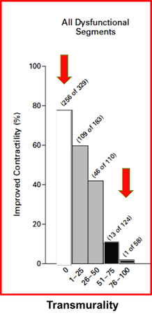
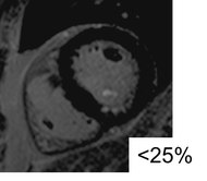
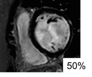
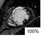
Clinical significance of transmurality of ischaemic scar in predicting improvement in contractile function with coronary revascularization. From Kim et al 2000

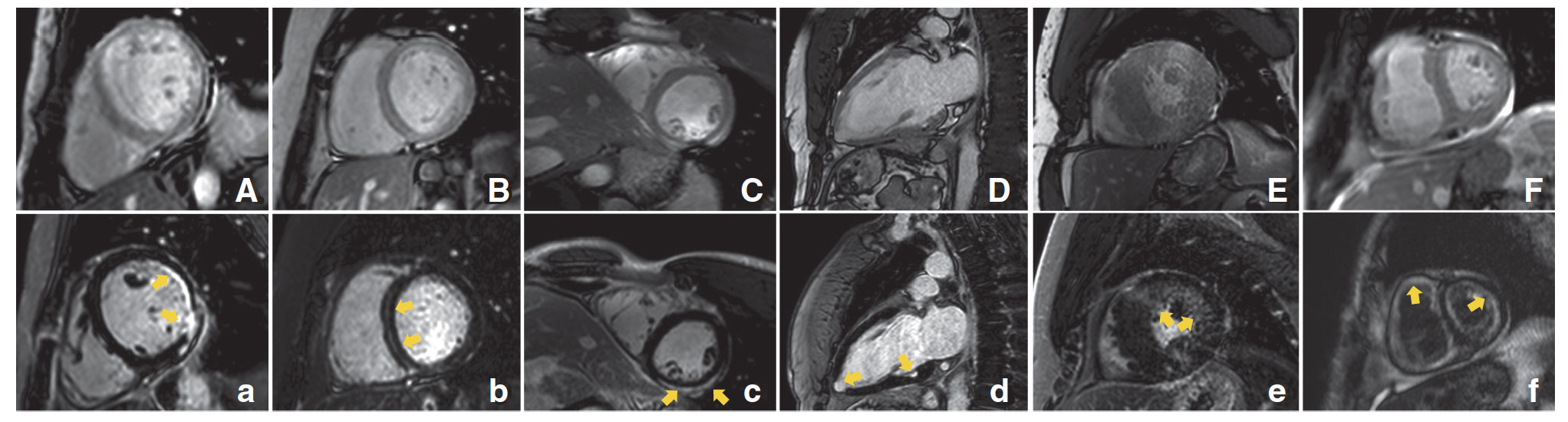
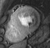
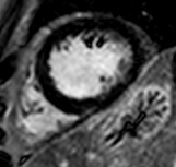
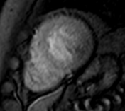
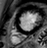
LGE patters in deciphering underlying aetiology in HF. A, Ischemic cardiomyopathy with subendocardial LGE in the antero- and inferolateral wall. B, Idiopathic dilated cardiomyopathy with mid-myocardial striae of LGE. C, Acute myocarditis with subepicardial LGE in the infero-lateral wall. D, Cardiac sarcoid with multiple focal areas of LGE (2CH view, particularly, in the inferior wall and apex). E, Hypertrophic cardiomyopathy with diffuse and circumferential LGE. F, Cardiac amyloid with diffuse and subendocardial enhancement. From: Puntmann et al. 2016
Picture

2. Chest pain - Angina Pectoris

1. Stable angina (chronic chest pain) 
stress-myocardial perfusion is the ultimate diagnostic test for the presence and relevance of myocardial ischaemia
  • to diagnose the presence myocardial ischaemia 
  • to guide management with revascularisation
​Guidelines recommendations: ESC Evidence level I (B), US: A

2. Patients presenting with ACS
  • assessment of AAR (salvageable myocardium) 
  • demonstration of microvascular obstruction (no-reflow phenomena) or intramyocardial hemorrhage -> poor prognostic markers
2. Patients presenting with ACS
  • assessment of AAR (salvageable myocardium) 
  • demonstration of microvascular obstruction (no-reflow phenomena) or intramyocardial hemorrhage -> poor prognostic markers
3. ACS-like symptoms with unobstructed coronary arteries
  • to confirm the ischaemic event 
  • to confirm the non-ischaemic aetiology: Tako-Tsubo, myocarditis, embolic events
Picture
MVO (no-reflow phenomenon)
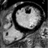
Adenosine stress test reveals a significant myocardial perfusion defect in basal-apical inferolateral segments. In the absence of myocardial scar in the LGE images, this finding speaks for relevant myocardial ischaemia. Left: adenosine perfusion, right: LGE.
Picture
Picture
Basal slice
Picture
Picture
Midventricular slice
Picture
Picture
Apical slice

3. Cardiomyopathies

Specific cardiomyopathies
1. Left ventricular hypertrophy
  • Hypertrophic Cardiomyopathy vs. hypertensive vs. amyloidosis, etc
2. Dilated cardiomyopathy
  • ? athletes heart vs. early stages  dilated cardiomyopathy vs. chronic myocarditis
  • risk stratification
3. Viral myocarditis
  • acute vs. chronic myocarditis
  • active chronic myocarditis
4. Cardiotoxicity
  • pre-chemotherapy risk assessment 
  • monitoring of anthracycline-chemotherapy, Herceptin
5. Systemic inflammatory conditions - Lupus, Sarcoidosis, Psoriasis, Rheumatoid arthritis, Systemic Sclerosis  
  • ? myocardial involvement;
  • monitoring of myocardial inflammation
  • risk of DCM or HF 
6. SCD survivors and Family screening:
  • survivors of SCD
  • relatives of patients with either known cardiomyopathies (HCM, ARVC, LVNC) or SCD
7. HFpEF Syndrome
8. 
Suspected infiltrative disease (?amyloidosis)
9. Patients at risk of cardiac iron loading (chronic transfusions)
10. Right ventricular enlargement
  • ?congenital heart disease,
  • pulmonary hypertension (?RV strain),
  • ? ARVC
11. Unclear aetiology of ECG changes, troponin rise, reduced LVEF
​Guidelines recommendations: ESC Evidence level II (B), US: A
Picture
Picture
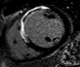
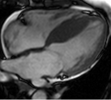
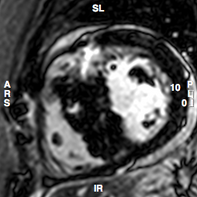
Hypertrophic cardiomyopathy. A. Cine 4CH view: asymmetric septal hypertrophy. B. LGE: evidence of scar in superior RV insertion point, From Puntmann et al. 2013.
Picture
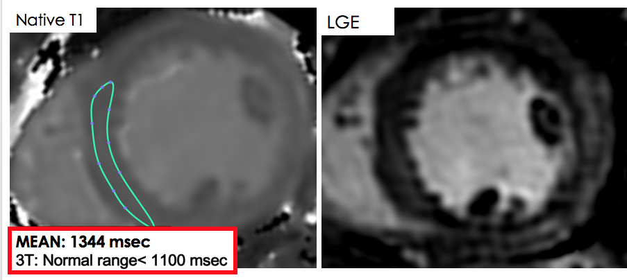
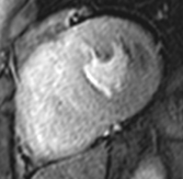
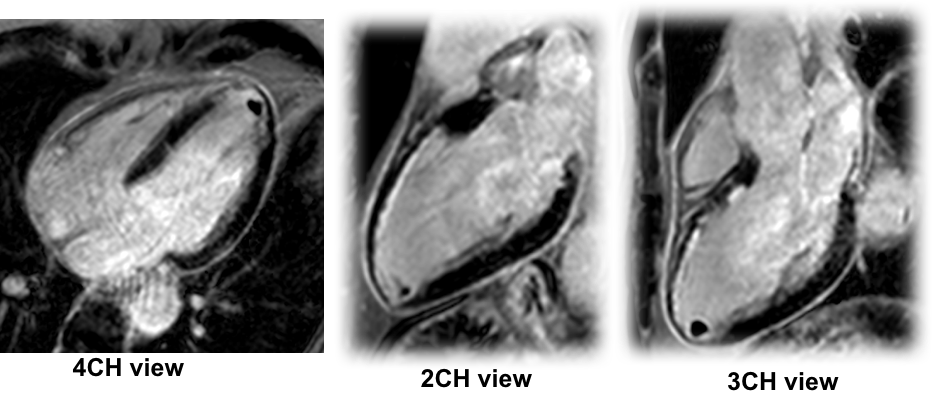
Acute myocarditis. A. Native T1 is highly elevated (>5SD). There is only a hint of diffuse LGE in the inferolateral segments. From Hinojar et al 2015
Picture
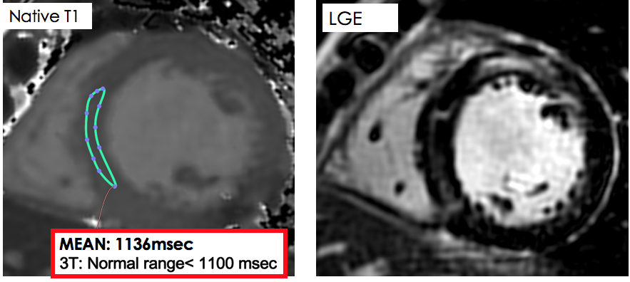
Chronic myocarditis. A. Native T1 is only mildly abnormal (>2SD). There is residual non-ischaemic intramyocardial scar in the anterior segments.
Picture
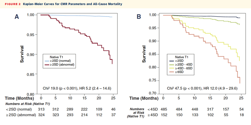
Native T1 and all-cause mortality in non-ischaemic dilated cardiomyopathy. From Puntmann et al. 2016

4. Cardiac Masses and Thrombi

Picture

5. Pericardial disease

1. Pericardial effusion
2. Pericarditis
  • acute perimyocarditis
  • chronic percarditis - monitoring of anti-inflammatory treatment
​
A- appropriate (US appropriateness criteria); I - level of recommendation for an indication (ESC guidelines)

6. Valvular disease

1. Aortic stenosis:
  • confirmation of aetiology of LVH (increased LV wall stress vs amyloid)
  • presence of fibrosis or scar
  • ? concomitant aortic disease (coarctation, dilatation)
2. Aortic regurgitation, Mitral Regurgitation
  • accurate determination of LV volumes 
  • mechanisms of valvular insufficiency (ischaemic vs functional) 
Overview on CMR indications in ESC guidelines (Von Knobelsdorff-Brenkenhoff F et al, JCMR 2016).
Powered by Create your own unique website with customizable templates.