Patients: HowTo CMR @ KGUWe offer high-end cardiovascular imaging within the Interdisciplinary Cardiovascular Imaging service provided in close collaboration with the Departments of Cardiology and Radiology, University Hospital Frankfurt. The scanning locations include Radiology Department (Haus 23C UG) and Institute for Experimental and Translational Cardiovascular Imaging (Haus 25B EG). All patients will be approached to take part in our research studies.
This webpage provides information specifically for the patients. For doctors, please click here.
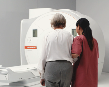
Courtesy of Siemens Healthineers.
How does it work?The Coordinating Team
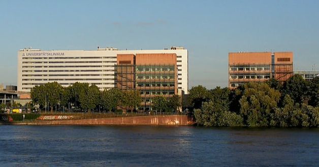
Where to find usHaus 25B Erdgeschoss, Theodor Stern Kai 7, 60590 Frankfurt am Main; Universitaetsklinikum, Institut fuer Experimentelle und Translationale Kardiovaskulaere Bildgebung; GoogleMap Coordinates
|
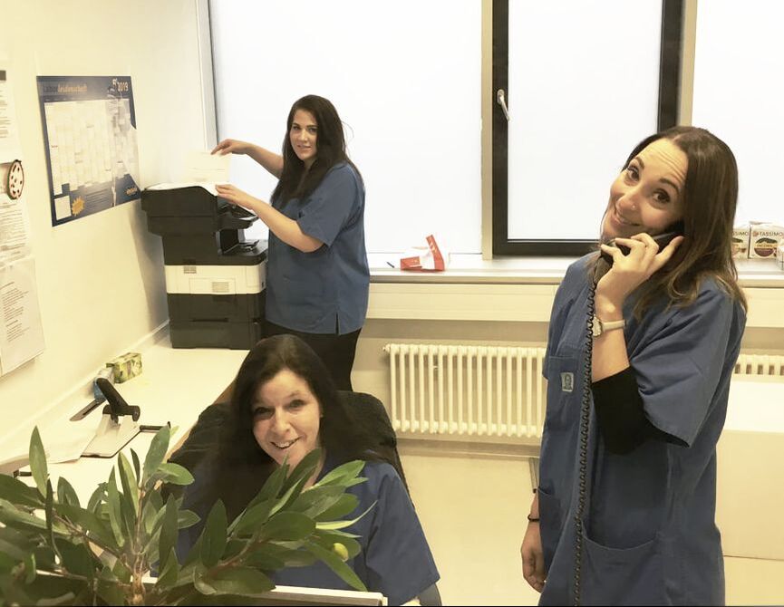
On the Day/Appointment
Patients will be asked to present to the Reception located on the ground floor of Haus 25B. Patients will be asked to fill out a short Health questionnaire. They will be the shown to the Scanning rooms (H25B or H23C). They will be asked to change in the hospital gown, to remove any jewellery, piercing, etc. We will have blood pressure measured and prepared for the investigation (IV access). This includes a chat with the Doctor about the examination as well as research study. Any remaining questions will be clarified. Patients will be asked to sign the consent forms.
The CMR Scan
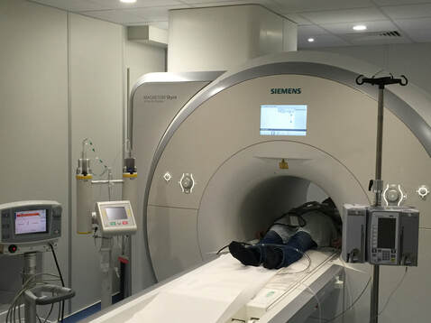
Patients will be shown to the MRI scanner room and further prepared for the scan. We will ask you to lie down on your back (supine), with the head towards the MRI scanner. We will place a sandbag between your legs to avoid any skin-to-skin contact and to remind to you to refrain from crossing your legs. ECG dots will be put on your chest and a blood pressure cuff will be placed on your upper arm. You will get an emergency bell, as well as headphones for noise protection. Finally we will place an antenna onto your chest below the chin, allowing the scanner to take images from the heart. After the preparation, you will slowly be driven into the centre of the scanner. Our scanner has a large bore with a diameter of 70 cm and is open at the front and the back. During the examination, various measurements are made in the entire area of the heart, as well as the outgoing vessels. It is important that you do not move the upper body during the entire measuring time. Please let us if we can do anything to make the procedure more comfortable for you. Depending on the question, the examination takes 20 to 50 minutes. During the entire examination, we offer all patients relaxation music via headphones. The measurements are ECG controlled and performed during breath-holds lasting about 10 to 20 seconds. Please let us know during the investigation if the breath holds are too long. In many cases, it is necessary to give a contrast agent, often together with stress-medication (Regadenoson). These enable us to evaluate the presence of the scar in the heart muscle, as well as the heart's blood flow. You will receive detailed information about these medications in the preparation material, as well as while providing informed consent with a doctor prior to the scan.
Findings and clinical report
A doctor performing the scan will provide you with a quick summary of the main findings immediately after the scan, however, all measurements and the final conclusions, including implications for future treatment, will be summarised in the clinical report. This will be send to your referring doctor (e.g. via internal electronic patient record system). It is important that you have secured a subsequent appointment in Cardiology outpatients to discuss the results and any possible changes to your treatment.
Clinical Research
All patients will be approached to take part in our research studies, in line with inclusion and exclusion criteria. Patients will be provided with the information and study material prior to the appointment. Most patients will be asked to take part in the Registry studies (International T1 Outcome Study, True-TypeCKD Study and Carysma Studies). In short, the participation in these registries includes obtaining patients' permission to reuse images of the clinical scans, undertake blood sampling and conduct telephonic follow-up in 2-5 years. There will be no additional procedures, drugs or contrast agents. Any questions will be clarified in a conversation with the imaging doctor on the day of your appointment. Any burning issues can also be addressed by sending an email to the coordinating team (email: cvi-info(at)kgu.de).
